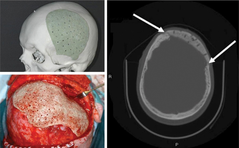
Fig. 2.

Additive manufacturing model of a large left calvarial bone defect with a tailor-made PMMA and B-G (S53P4) implant before operation (above, left). Intraoperative picture of a bioactive composite implant adjusted to its correct position in the calvarial bone defect. Note the 1.5 mm perforations to enhance tissue growth into the alloplastic material (below, left). CT scan of a left temporal bone defect 2 years after reconstruction (right). Implant is in the correct position in the skull. New bone formation between implant and surrounding bone is seen (white arrows). CT, computed tomography; PMMA, polymethylmethacrylate. (Adapted from Peltola et al.30)
