. 2015 Dec 17;4:e08887. doi: 10.7554/eLife.08887

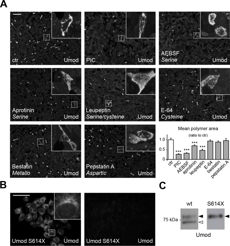
Figure 2. A serine protease is responsible for the release of polymerisation-competent uromodulin.

(A) Immunofluorescence analysis showing uromodulin on the surface of MDCK cells treated with vehicle (DMSO) (ctr), protease inhibitor cocktail (PIC) or single PIC components, as indicated. Scale bar, 50 µm. Quantification of the average surface of uromodulin polymers shows that only treatment with PIC or with specific serine protease inhibitors (AEBSF, aprotinin and leupeptin) significantly reduces uromodulin polymerisation on the surface of MDCK cells. Bars indicate average ± s.e.m. ***p<0.001 (Mann-Whitney test). The graph represents mean ratios of 3 independent experiments (Figure 2—source data 1). (B) Immunofluorescence analysis of permeabilised (left) or non-permeabilised (right) MDCK cells expressing soluble uromodulin mutant S614X. Uromodulin polymerisation is abolished by preventing its association to the membrane. Scale bar, 50 µm. (C) Representative Western blot analysis of N-deglycosylated uromodulin secreted by MDCK cells expressing wild-type or soluble (S614X) uromodulin. Lack of plasma membrane anchoring does not affect uromodulin secretion but it abolishes its cleavage at the physiological site (white arrowhead).

DOI: http://dx.doi.org/10.7554/eLife.08887.004

Figure 2—source data 1. Quantification of the area of uromodulin polymers on the surface of MDCK cells after protease inhibitor treatment (Figure 2A).
DOI: 10.7554/eLife.08887.005
Figure 2—source data 2. Short cleavage inhibition by HAI-1 expression (Figure 2—figure supplement 2C).
DOI: 10.7554/eLife.08887.006

Figure 2.

Figure 2—figure supplement 1. PIC treatment does not affect uromodulin intracellular distribution and expression.
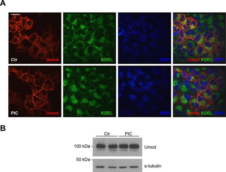
Figure 2—figure supplement 1.

(A) Immunofluorescence analysis showing intracellular distribution of uromodulin in MDCK cells treated with vehicle (DMSO) (ctr) or protease inhibitor cocktail (PIC). KDEL is a marker of the endoplasmic reticulum (ER). Scale bar, 16 µm. (B) Representative Western blot analysis of uromodulin in lysates of MDCK cells under the same conditions as above. The upper band corresponds to the mature, fully glycosylated protein, the lower band corresponds to the immature protein carrying ER-type glycosylation (Schaeffer et al., 2012). Alpha-tubulin is shown as a loading control. PIC treatment does not alter uromodulin intracellular distribution nor its expression.
Figure 2—figure supplement 2. Expression of the serine protease inhibitor HAI-1 effectively reduces uromodulin cleavage at the urinary site.
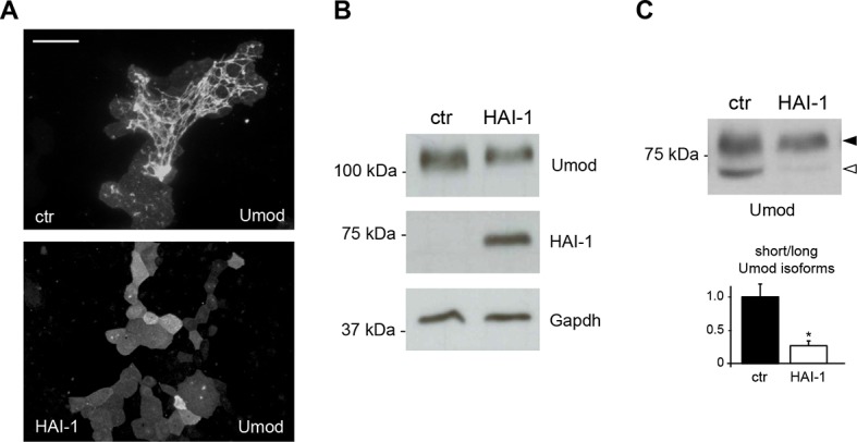
Figure 2—figure supplement 2.

(A) Immunofluorescence analysis showing uromodulin on the surface of MDCK cells co-expressing the serine protease inhibitor HAI-1 (Hepatocyte growth factor Activator Inhibitor-1), as indicated. HAI-1 expression essentially abolishes uromodulin polymerisation on the cell surface. Scale bar, 50 µm. (B) Representative Western blot analysis showing uromodulin and HAI-1 expression in cell lysates of transfected MDCK cells, as indicated. Glyceraldehyde-3-phosphate dehydrogenase (Gapdh) is shown as a loading control. (C) Representative Western blot analysis of N-deglycosylated uromodulin secreted by MDCK cells co-expressing HAI-1, as indicated. Densitometric analysis (average ± s.d. of 3 independent experiments, Figure 2—source data 2) shows the ratio between the short and the long uromodulin isoforms in the absence or presence of HAI-1 co-expression, as indicated. The serine protease inhibitor HAI-1 strongly reduces the amount of the short uromodulin isoform released in the culturing medium by MDCK cells. *p<0.05 (Student’s t test).