. 2015 Dec 9;4:e10766. doi: 10.7554/eLife.10766

Figure 1. Residues 614-682 of CD11b are crucial for CyaA binding and penetration into cells.

(A) The CD11 subunits of β2 integrins consist of a long N-terminal extracellular domain, a single-pass transmembrane segment (TS) and a short C-terminal cytoplasmic tail, respectively. The N-terminal part of the extracellular domain harbors seven β-sheet repeats (numbers in boxes), forming a β-propeller domain, which is followed by the thigh, calf-1 and calf-2 domains. The I-domain segment, inserted between repeats 2 and 3 of the β-propeller domain, plays a critical role in interaction of the I-domain-containing integrins with their endogenous ligands. To map the CyaA binding site on the CD11b subunit, segments of CD11b (green) were systematically replaced with their CD11c counterparts (blue). In the CD11bΔ164-339 molecule, the entire I domain of CD11b was deleted. (B) 2x10⁵ CHO cells expressing integrin molecules were incubated with 2 µg/ml of CyaA-biotin, the surface-bound toxin was labeled with streptavidin-PE and the cells were analyzed by flow cytometry. CyaA binding was expressed as percentage of toxin binding to CHO cells expressing the native form of CD11b/CD18. Each bar represents the mean value with SD of at least five independent experiments performed in duplicate or triplicate. Significantly reduced binding of CyaA to mutant integrins in comparison with intact CD11b/CD18 is indicated (****, p<0.0001; ANOVA). (C) 1x10⁵ CHO cells expressing integrin molecules were incubated with various concentrations of CyaA and the amounts of accumulated cAMP were determined in cell lysates by ELISA. Each point represents the mean value ± SD of at least seven determinations from at least three independent experiments. Significant differences between mean values of cAMP intoxication of cells expressing intact CD11b/CD18 and mutant integrins are shown (****, p<0.0001; ANOVA).

DOI: http://dx.doi.org/10.7554/eLife.10766.003

Figure 1.

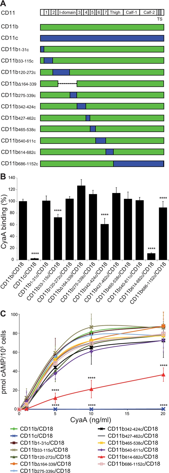
Figure 1—figure supplement 1. Expression of the CD18 subunit on the surface of CHO cells.

Figure 1—figure supplement 1.

A CHO cell line was transfected with a plasmid construct encoding CD18 and the positively transfected cells were selected using a cell sorter. The expression of the CD18 subunit (violet) on the cell surface was examined by flow cytometry upon staining of 2x10⁵ cells with the anti-CD18 mAb MEM-48. CHO cells transfected with an empty vector were processed in parallel and used as negative control (grey). A typical flow cytometry histogram from one representative binding experiment out of five performed is given. RFI, relative fluorescence intensity.
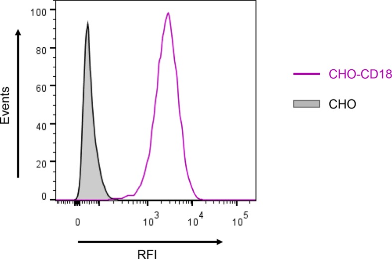
Figure 1—figure supplement 2. Binding of mAbs and CyaA to CHO cells expressing CD11b/CD18, CD11c/CD18 and the CD11b-CD11c/CD18 chimeras.

Figure 1—figure supplement 2.

A CHO cell line stably expressing intact CD18 subunit was transfected with plasmid constructs encoding CD11b, CD11c, or their respective chimeric variants. Cells stably expressing the integrin molecules were selected using a cell sorter. The expression levels of CD11b/CD18 (green), CD11c/CD18 (blue) and CD11b-CD11c/CD18 chimeras (red) on the cell surface were examined by flow cytometry upon staining of 2x10⁵ cells with the ICRF 44 mAb (recognizing the I-domain of CD11b (left panels)), the OKM1 mAb (recognizing segment 614-682 of CD11b (middle left panels)), or the 3.9 mAb (recognizing the I-domain of CD11c (middle right panels)). The cells were also analyzed for capacity to bind CyaA: 2x10⁵ transfected cells were incubated with 2 μg/ml of CyaA-biotin, the surface-bound toxin was labeled with streptavidin-PE and the cells were analyzed by flow cytometry (right panels). A typical histogram from one representative binding experiment out of five performed is shown for binding of the respective mAb (ICRF 44, OKM1 or 3.9), or CyaA to each integrin variant. CHO cells expressing no β2 integrin (transfected with empty vectors) were processed in parallel and used as negative control (grey). RFI, relative fluorescence intensity.
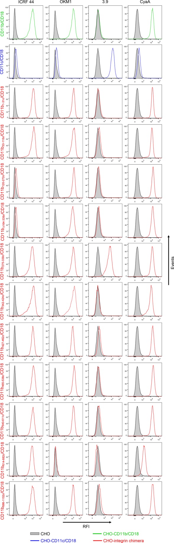
Figure 1—figure supplement 3. CyaA recognizes CD11b only in the heterodimeric complex with CD18.

Figure 1—figure supplement 3.

(A and B) 1x10⁵ CHO cells stably expressing intact dimeric CD11b/CD18 or its individual monomeric subunits CD11b or CD18 were stained with the anti-CD11b mAb OKM1 (A) or the anti-CD18 mAb MEM-48 (B) and analyzed by flow cytometry. A typical overlay flow cytometry histogram from one representative binding experiment out of three performed is given. (C) 1x10⁵ integrin-transfected CHO cells were incubated with different concentrations of Dy647-labeled CyaA and the cells were analyzed by flow cytometry. CyaA binding data were deduced from the mean fluorescence intensities of three independent experiments and each point represents the mean value ± SD. Binding of CyaA to cells expressing CD11b, CD18 or no β2 integrin was at all measured toxin concentrations significantly lower than toxin binding to cells expressing intact CD11b/CD18 (****, p<0.0001; ANOVA). CHO cells transfected with an empty vector and expressing no β2 integrin were processed in parallel and used as negative control (all panels). RFI, relative fluorescence intensity.