. 2015 Dec 9;4:e10766. doi: 10.7554/eLife.10766

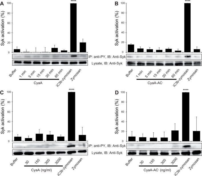
Figure 6. CyaA binding to CR3 does not trigger Syk activation.

(A, B) 3x10⁶ THP-1 cells were incubated with 30 ng/ml of CyaA (A), or CyaA-AC- (B) for indicated times. (C, D) 3x10⁶ THP-1 cells were incubated with different indicated concentrations of CyaA for 15 min (C), or CyaA-AC- for 30 min (D). (A-D) Treated cells were lysed and cell lysates were immunoprecipitated (IP) with anti-phosphotyrosine (anti-PY) mAb. Syk-P immunoprecipitated from whole cell lysates was detected by immunoblotting (IB) with anti-Syk mAb and normalized to total Syk detected in whole cell lysates. Cells treated with iC3b-opsonized zymosan were taken as a positive control and the cells treated with buffer, or unopsonized zymosan were taken as negative controls. Each bar represents the mean value with SD of three independent experiments. In comparison to buffer-treated cells, a significant increase of Syk activation was observed only in cells treated with iC3b-opsonized zymosan (****, p<0.0001; ANOVA).

DOI: http://dx.doi.org/10.7554/eLife.10766.019

Figure 6.

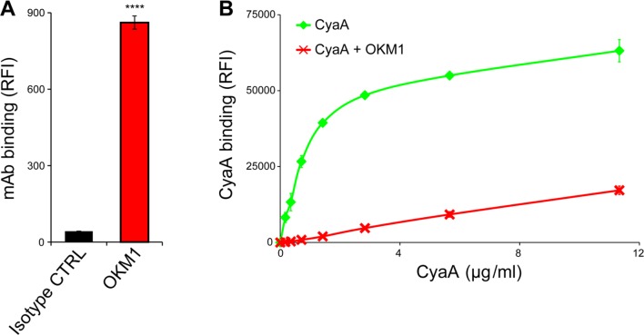
Figure 6—figure supplement 1. CyaA binds efficiently and specifically the THP-1 cells.

Figure 6—figure supplement 1.

(A) 2x10⁵ THP-1 cells were treated with the anti-CD11b OKM1 mAb, or with an isotype-matched mouse IgG2b control antibody and the cells were analyzed by flow cytometry. Each bar represents the mean value ± SD of two independent experiments performed in duplicate. Significant difference between mean values of OKM1 and the control mAb binding to THP-1 cells is indicated (****, P < 0.0001; Student’s t-test). (B) 2x10⁵ THP-1 cells were preincubated without or with 25 μg/ml of the anti-CD11b OKM1 mAb for 30 min and subsequently treated with different concentrations of CyaA-biotin. The surface-bound toxin was labeled with streptavidin-PE and the cells were analyzed by flow cytometry. Mean fluorescence intensities of CyaA binding were plotted against toxin concentrations. Each point represents the mean value ± SD of two independent experiments performed in duplicate. Binding of CyaA to cells preincubated with the OKM1 mAb was at all measured concentrations significantly lower than binding of CyaA to cells pre-treated with buffer alone (P < 0.01; Student’s t-test). RFI, relative fluorescence intensity.