. 2015 Dec 9;4:e10766. doi: 10.7554/eLife.10766

Figure 9. CyaA binds and intoxicates cells expressing LFA-1 with equally low efficacy as cells expressing CR4, or lacking any β2 integrin at all.

(A) 2x10⁵ stably transfected CHO cells expressing CD11a-YFP/CD18, CD11b-YFP/CD18, CD11c-YFP/CD18, or no β2 integrin were incubated with different concentrations of Dy647-labeled intact CyaA and analyzed by flow cytometry. Mean fluorescence intensities of CyaA binding were plotted against the concentrations of CyaA. RFI, relative fluorescence intensity. (B) 1x10⁵ CHO cells expressing integrin molecules were incubated at indicated concentrations of CyaA and the amounts of accumulated cAMP were determined in cell lysates by ELISA. (A and B) Each point represents the mean value ± SD of three independent experiments performed in duplicate. CyaA binding to or cAMP intoxication of cells expressing CD11a-YFP/CD18, CD11c-YFP/CD18, or no β2 integrin was at all toxin concentrations significantly lower than toxin binding to or cAMP intoxication of cells expressing intact CD11b-YFP/CD18 (p<0.0001; ANOVA). However, CyaA binding to or cAMP intoxication of cells expressing CD11a-YFP/CD18 was at all measured toxin concentrations found to be statistically the same as toxin binding to or cAMP intoxication of cells expressing CD11c-YFP/CD18 or no β2 integrin at all (P > 0.1; ANOVA).

DOI: http://dx.doi.org/10.7554/eLife.10766.029

Figure 9.

Figure 9—figure supplement 1. Expression of integrin variants fused with a fluorescent YFP protein on the surface of CHO cells.

Figure 9—figure supplement 1.

A CHO cell line stably expressing the intact CD18 subunit was transfected with plasmid constructs encoding CD11b-YFP or CD11a-YFP and cells stably expressing the integrin molecules were selected using a cell sorter. The expression levels of CD11b-YFP/CD18 (green) and CD11a-YFP/CD18 (violet) on the surface of 2x10⁵ cells were examined by flow cytometry for YFP (left panels), or upon staining of cells with a mAb recognizing CD11b (M1/70, middle panels), or a mAb recognizing CD11a (MEM-25, right panels). CHO cells expressing no β2 integrin (transfected with empty vectors) were used as negative control (grey). Typical flow cytometry histograms from one representative binding experiment out of four performed are shown. RFI, relative fluorescence intensity.

Figure 9—figure supplement 2. Residual binding of CyaA to CD11a-YFP/CD18 is not due to the presence of the YFP tag.

Figure 9—figure supplement 2.

(A) A CHO cell line stably expressing the intact CD18 subunit was transfected with a plasmid construct encoding CD11a and cells stably expressing CD11a/CD18 were selected using a cell sorter. The expression levels of CD11a/CD18 (red) on the surface of 2x10⁵ cells were examined by flow cytometry upon staining of cells with the anti-CD11a mAb MEM-25 (left panels) and compared with expression levels of CD11a-YFP/CD18 (violet). Both CD11a/CD18- and CD11a-YFP/CD18-epressing cells were also examined by flow cytometry for YFP (right panels). CHO cells expressing no β2 integrin (transfected with empty vectors) were used as negative control (grey). Typical flow cytometry histograms from one representative binding experiment out of four performed are shown. RFI, relative fluorescence intensity. (B) 2x10⁵ stably transfected CHO cells expressing CD11a/CD18, CD11a-YFP/CD18, CD11b/CD18, or no β2 integrin were incubated with different concentrations of Dy647-labeled intact CyaA and analyzed by flow cytometry. Mean fluorescence intensities of CyaA binding were plotted against the concentrations of CyaA. RFI, relative fluorescence intensity. Each point represents the mean value ± SD of two independent experiments performed in duplicate. CyaA binding to cells expressing CD11a/CD18, CD11a-YFP/CD18 or no β2 integrin was at all toxin concentrations significantly lower than toxin binding to cells expressing intact CD11b/CD18 (p<0.0001; ANOVA). In contrast, CyaA binding to cells expressing CD11a/CD18 at all measured toxin concentrations was found to be statistically the same as toxin binding to cells expressing CD11a-YFP/CD18 or no β2 integrin at all (P > 0.1; ANOVA).

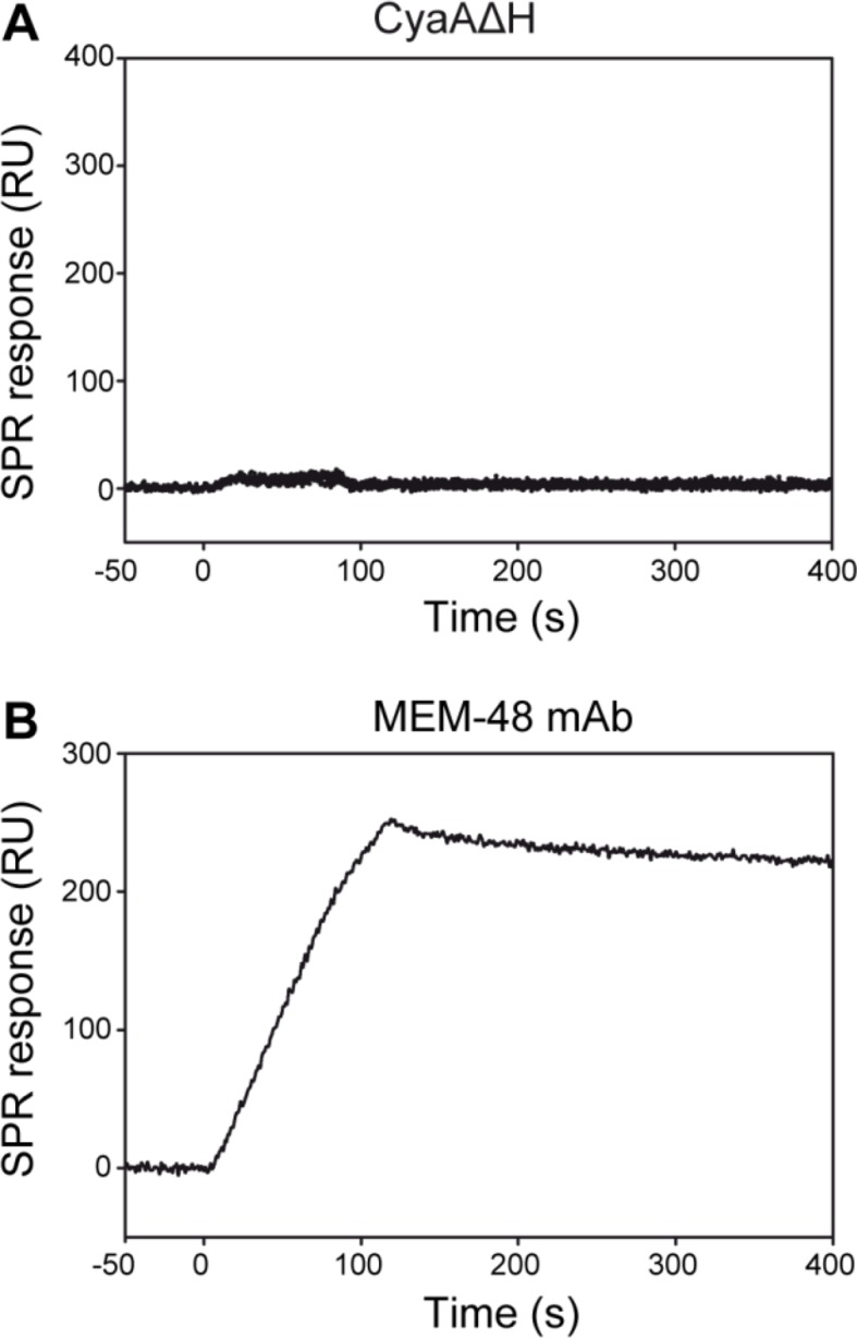
Figure 9—figure supplement 3. CyaA does not interact with the intact native LFA-1 integrin.

Figure 9—figure supplement 3.

(A) LFA-1 was purified from human peripheral blood mononuclear cells by immunoaffinity chromatography and immobilized onto an SPR sensor chip. To analyze the interaction between LFA-1 and the toxin, CyaA∆H was passed over the chip surface at concentrations of 20, 40, 80, 160 and 320 µg/ml. (B) The MEM-48 mAb recognizing the CD18 subunit of LFA-1 was used as positive control.