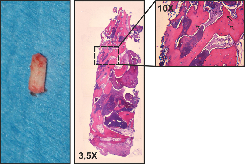
Fig. 5.

One of the two biopsies collected from the grafted site and the hematoxylin-eosin staining of the bioptic sample. The 10X magnification detail shows the absence of inflammatory reaction and the graft material undergoing remodeling; black arrows indicate the osteocytes into the newly formed bone.
