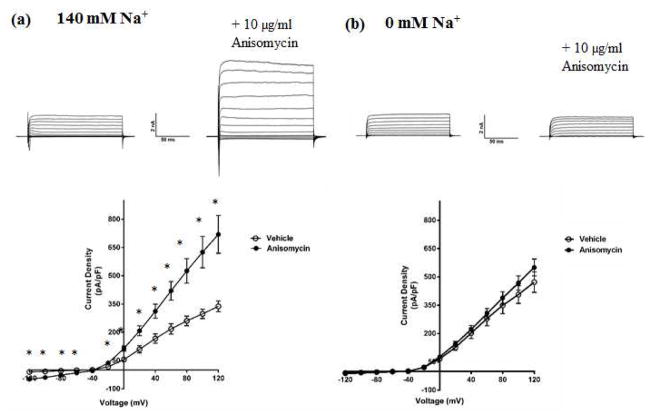
Fig. 2. Activation of p38 MAPK by anisomycin up-regulates the KNa component of IK in DRG neurons.
(a) Top- Representative traces from whole-cell recordings of DRG neurons treated with 10μg/ml anisomycin for 90 minutes at 37 °C, in bath solution containing sodium. Bottom- Current-Voltage relationship of IK measured from anisomycin-treated DRG neurons in bath solution containing sodium. (b) Top- Representative traces from whole-cell recordings of DRG neurons treated with 10μg/ml anisomycin for 90 minutes at 37 °C, in bath solution containing the impermeant cation NMG in place of sodium. Bottom, Current-Voltage relationship of the isolated KNa current measured from anisomycin-treated DRG neurons in bath solution containing NMG. Holding potential was −70 mV, and currents were elicited with voltage steps from −120 mV to +120 mV in 20 mV increments. Data are means ± SEM (n=8–12 per group). Statistical analysis was done using Student’s unpaired t-test (*p<0.05).

