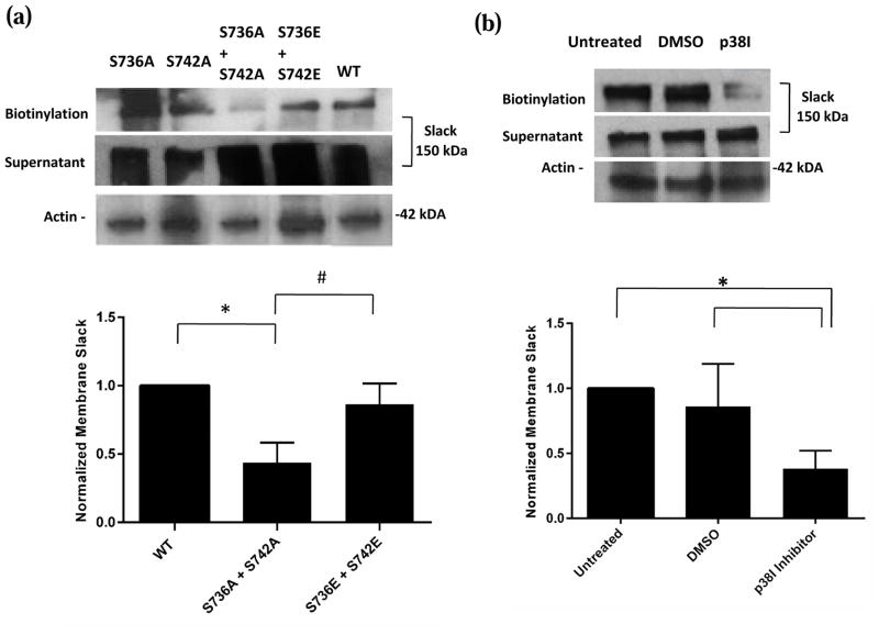
Fig. 7. p38 MAPK phosphorylation at Serines 736 and 742 is required for surface expression of Slack.
(a) Top- Representative images of western blots for membrane Slack following biotinylation assays on lysates of CHO cells transiently transfected with WT, double alanine mutant or double glutamate mutant Slack pTRACER. Actin was used as a loading control. Bottom- Densitometric analysis of represented western blots for Slack quantified as normalized to Actin. Data is expressed as mean +/− SEM (n=4 for all groups). Statistics performed using One Way ANOVA followed by multiple comparisons using Tukey’s method (*p<0.05 between WT and S736A+S742A groups, #p<0.05 between S736E+S742E and S736A+S742A groups). (b) Top- Representative images of western blots for membrane Slack following biotinylation assays on lysates of Slack stable HEK cells incubated in Vehicle (0.01% DMSO) or p38 MAPK Inhibitor (10 μM) for 90 minutes at 37 °C. Actin was used as a loading control. Bottom- Densitometric analysis of represented western blots for Slack quantified as normalized to Actin. Data is expressed as mean +/− SEM (n=3 for all groups). Statistics performed using One Way ANOVA followed by multiple comparisons using Tukey’s method (*p<0.05).

