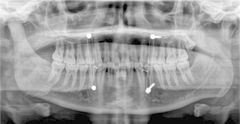
Fig. 3.

Panoramic radiograph of patient in Case 1 featured immediately postoperatively in maxillomandibular fixation. Note the lack of any fixation of the spacer as well as the excellent symmetry and condyle/fossa relationship when compared with the healthy condyle.
