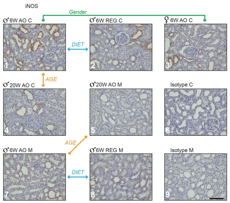
Figure 6.
iNOS positive staining in obese Zucker rat kidney cortex and medulla. Panels 1 through 7: Zucker rat kidney stained immune-histochemically for iNOS; representative samples are shown from groups demonstrating statistically significant differences in staining. Panels 8 and 9 show representative Zucker rat kidney sections treated with non-immune serum (isotype control). M-medulla; C-cortex; 6 w- 6 weeks, 20 w- 20 weeks. Magnification: 200×, bar = 100μm.

