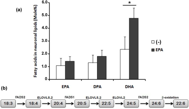
Figure 5. Addition of EPA increased docosahexaenoic acid (DHA) levels in retina neurons.

Retina neurons were supplemented with vehicle (−) or with 3 μM EPA added at day 1 in vitro and the fatty acid composition of neuronal phospholipids was determined by GLC at day 4. Bars (a) depict mean ± SD of the mole % content of EPA, docosapentaenoic acid (DPA) and DHA in neuronal phospholipids. Three different experiments with two dishes for each condition were analyzed. * p≤0.05, statistically significant differences, compared to the respective controls, determined by a Student’s t-test. Sequence of metabolic reactions (b) leading to 22:6 n-3 (DHA) synthesis from 18:3 n-3 that involves its desaturation by FADS2, the further elongation and desaturation catalyzed by ELOVL5,2 and FADS1 to synthesize 20:5 n-3 (EPA), which is successively elongated by ELOVL5,2 and ELOVL2 to 24:5 n-3, which is then desaturated by FADS2 to 24:6 n-3, which after a (peroxisomal) β-oxidation gives rise to DHA.
