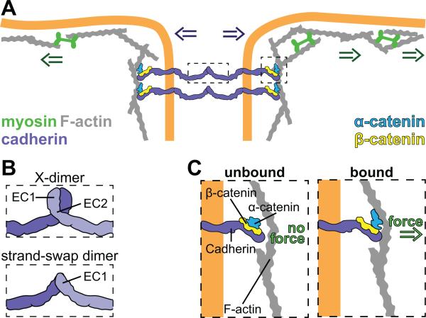
Figure 1. Adherens junctions mechanically couple adjacent cells.
(A) AJs are composed of cadherins, α-catenin, and β-catenin. The extracellular domains of cadherins can interact with each other to link adjacent cells. Together α-catenin and β-catenin form a scaffold that links the AJ to the actin cytoskeleton. In this way the intracellular force from actomyosin contraction (green arrows) is transmitted to adjacent cells (purple arrows). (B) Schematic of two different conformations for trans-interaction of vertebrate E-Cadherin highlighting the regions of interest. E-Cadherin can adopt the X-dimer form (top) or the strand-swapped dimer form (bottom). (C) Components of the AJs complex interact in a force dependent manner. β-catenin binds directly to the intracellular domain of E-cadherin in the presence or absence of force. In the absence of force, α-catenin adopts a folded conformation and does not bind strongly to actin filaments (F-actin) (left). When tensile force is applied to the junction, α-catenin adopts a new conformation and binds F-actin tightly (right).

