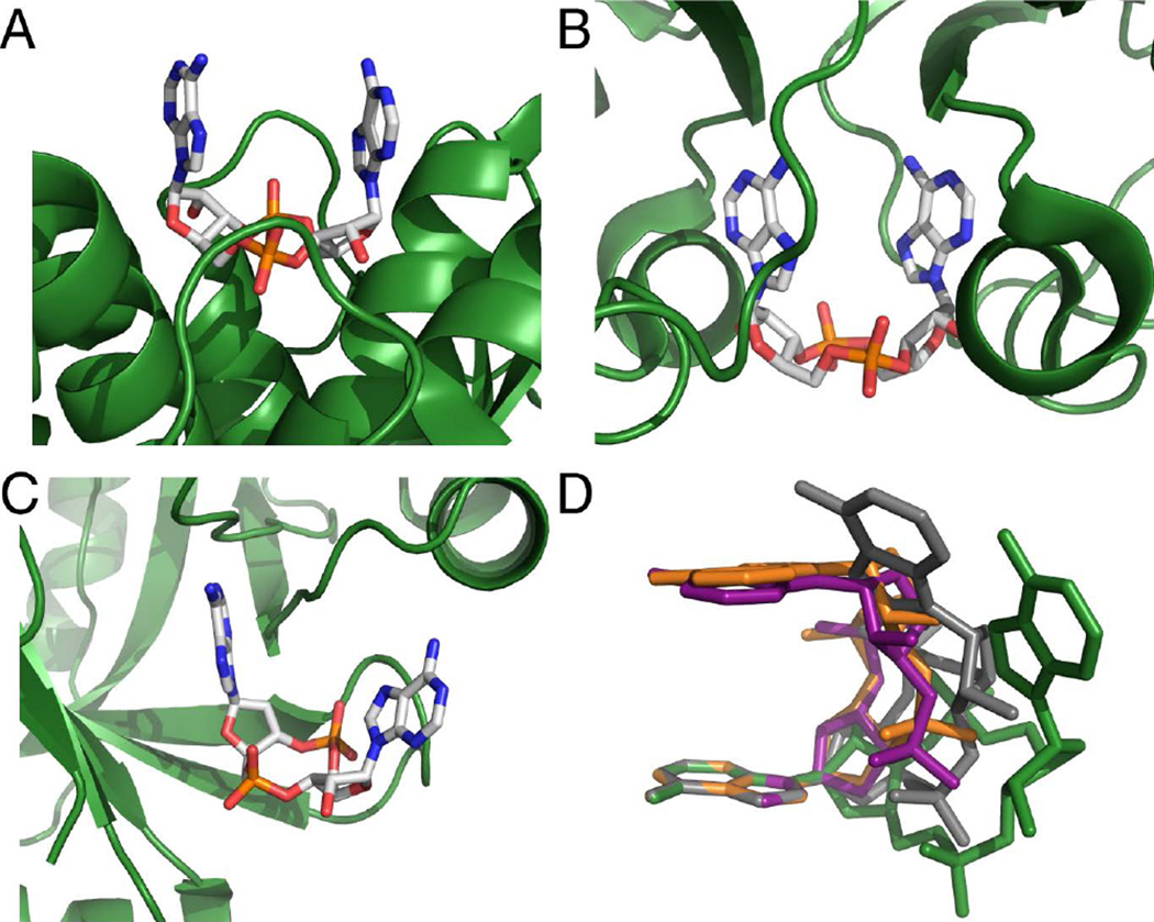
Figure 1.
c-di-AMP bound to macromolecular receptors (A) LmPC, (B) KtrA, and (C) PstASA (ref. 32,33,37). c-di-AMP is colored by atom with carbon in white, oxygen in red, nitrogen in blue, and phosphorous in orange. Protein binding sites are shown as green cartoons. (D) Conformations adopted by c-di-AMP when bound to downstream targets (ref. 32,33,37,45–47). C-di-AMP is colored in green for ydaO, grey for PstASA, orange for LmPC, and purple for KtrA. The structures are superimposed at a single adenosine, with the remaining atoms floating.

