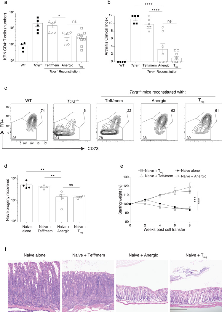
Figure 6.
Treg cells generated from anergic CD4+ polyclonal T cells prevent arthritis and colitis. (a–c) Lymphopenic Tcra−/− B6G7F1 mice were reconstituted with 1.5 × 10⁶ sorted syngeneic Teff/mem, anergic, or Treg cell CD4+ polyclonal T cells from a Foxp3DTR mouse for a period of 21 d. On day 21, 104 naive KRN transgenic CD4+ T cells were transferred into these mice (as well as into control WT and Tcra−/− B6G7F1 hosts) and then recipients were monitored over 12 d for arthritis development as described9, with the (a) number of KRN cells recovered, and (b) clinical arthritis index score on day 12 after KRN cell transfer as indicated. (c) CD73 and FR4 expression on KRN T cells on day 33. (d–f) A total of 2 × 10⁵ congenic–marked naive polyclonal CD4+ T cells were adoptively transferred into Tcra−/− B6 mice either alone or in combination with 4 × 10⁵ Teff/mem, anergic, or Treg cell CD4+ polyclonal cells sorted from Foxp3GFP mice, and then recipients were monitored over 8 weeks for weight loss. (d) Number of donor naive cells recovered at the end of week 8. (e) Change in body weight of Tcra−/− B6 mice receiving naive cells alone, or naive plus Teff/mem, anergic or Treg cells. (f) Representative H&E staining of the colon (all panels same magnification, 200µm). Mean data shown are representative of 3 (a–c) or 2 (d–f) independent experiments, with n = 2 to 3 mice per group. Error bars represent the SEM. (a–c, d). Cells pooled from spleen and lymph nodes (inguinal, axillary, brachial, cervical, mesenteric and pancreatic). One-way ANOVA; * p < 0.05, ** p < 0.01, *** p < 0.001 **** p < 0.0001, non-significant (ns). Points denote individual mice.

