Figure 6.
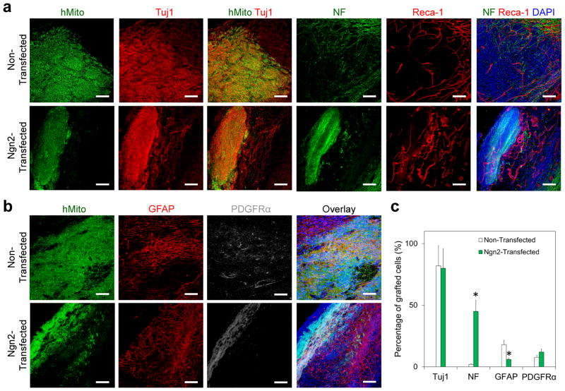
Mature neuron derived from transplanted Ngn2-transfected hNSCs at the brain lesion site. (a, b) Immunofluorescence staining confirmed the differentiation of transplanted hNSCs into (a) neurons [Tuj1+ and neurofilament (NF)+, red] and (b) glial cells (GFAP+, red, and PDGFRα+, gray) at 4 weeks after transplantation with our tailored HA hydrogel. Transplanted cells were identified by human mitochondria with green. DAPI was stained for cell nuclei with blue. (c) Quantitative analysis of differentiation of transplanted hNSCs at the lesion site. Ngn2-transfected hNSCs generated significantly more mature neurons (NF+) and less astrocytes (GFAP+) than non-transfected cells at the lesion site at 4 weeks after transplantation (n = 3; *P < 0.05). Scale bar = 100 μm.

