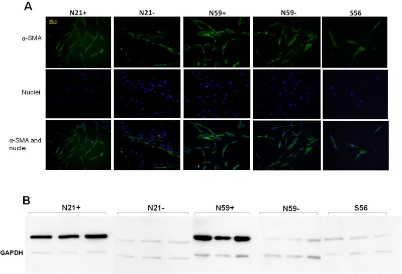
Figure 4.
(A) Images by immunocytochemistry demonstrating α-SMA expression in N21+, N21−, N59+, N59− and S56 VFF. N21+ and N59+ VFF appear to have enhanced of α-SMA expression. Nuclei were stained with 4',6-diamidino-2-phenylindole (DAPI). (B) Western blot images showed evident difference of α-SMA expression in N21+ and N59+ compared to N21−, N59− and S56 VFF versus loading control (GAPDH).

