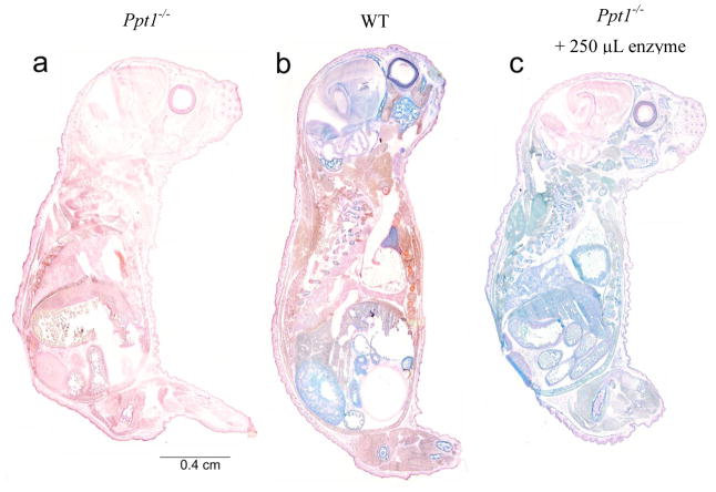
Figure 2. Histochemical stain for PPT1 activity in newborn mice.
Sagittal sections of entire newborn (PND1) mice were stained for PPT1 activity (blue) and counterstained with nuclear fast red. PPT1 activity is undetectable in untreated Ppt1−/− mice (a) and is indicated by discrete regions of intense blue in WT mice (b). Staining of tissue in enzyme-treated Ppt1−/− mice (c) is more uniformly blue, with the exception of the brain, indicating the presence of PPT1 activity. There also appears to be less intense staining in the retina of enzyme- treated animals compared to WT.

