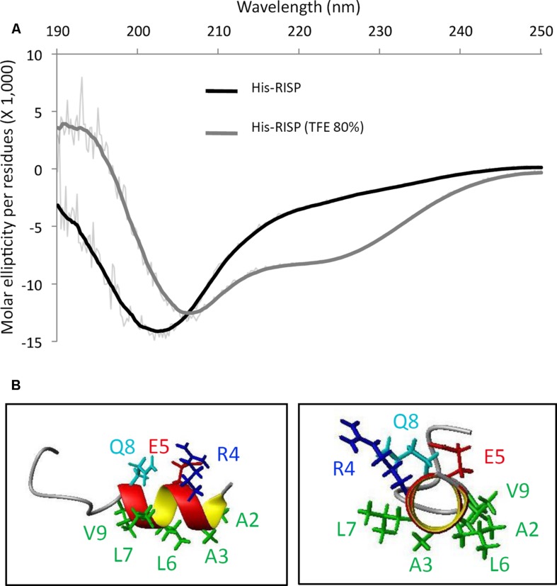
FIGURE 2.
RISP is intrinsically disordered and adopts α-helices in TFE. (A) Circular dichroism (CD) spectra of His-RISP in aqueous solution (phosphate buffer pH 7.0, in black) or in 80% trifluoroethanol (TFE; in gray). Thick lines: smoothed curves (n = 25), thin lines: curves established with raw CD data. (B) Nuclear magnetic resonance (NMR) model of the 15 N-terminal residues of His-RISP in 80% TFE-d3/20% phosphate buffer pH 7.4, at 25°C. Side chains of residues 2–9 are represented in green (hydrophobic side chains), dark blue (positively charged side chain), red (negatively charged side chain) or light blue (polar side chain).

