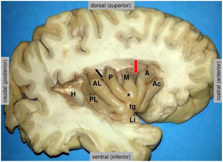
Figure 2.
Anatomy of the insula, as disclosed in the depth of the lateral fissure. H = the posterior medial stub of the transverse temporal gyrus of Heschl (primary auditory cortex) that was resected to uncover the posterior long insular gyrus. The central sulcus (black arrow) divides the lateral surface of the insula into a small posterior insular lobule, composed of the anterior long (AL) and posterior long (PL) insular gyri that converge to the limen insulae (Li), and a large anterior insular lobule, composed of the anterior short (A), the middle short (M) and the posterior short (P) insular gyri that converge to the apex of the insula (*). The anterior face of the insula displays a variably present accessory insular gyrus (Ac) and a constant transverse insular gyrus (tg) that connects with the orbital surface of the frontal lobe. The red arrow marks the sulcus between the anterior short and middle short gyri, where the functional “overlap region” was found by Kurth et al. (2010). Figure courtesy of Drs. Thomas P. Naidich and Mary E. Fowkes, the Icahn School of Medicine at Mt. Sinai, New York.

