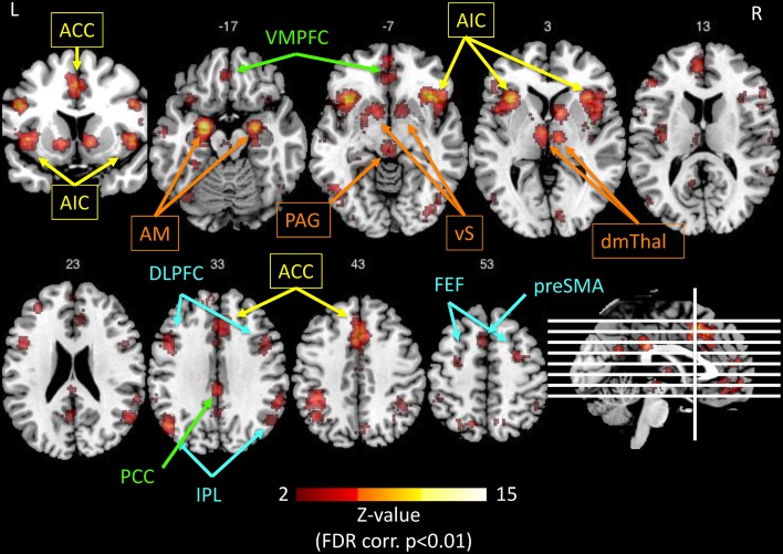
Figure 4.
Illustration of the networks implicated in salience processing, as obtained by the meta-analysis tool Neurosynth.org (Yarkoni et al., 2011), based on the results of 122 published studies, using the only keyword “salience” in forward inference, and displayed on a template anatomical T1 image with Z-values of 2 to 15 (using the Mango software package2). The resulting significant regions (p < 0.01, FDR corrected) interestingly comprise four networks: Yellow: anterior cingulate cortex (ACC) and anterior insula (AI) represent the salience network (Seeley et al., 2007b). Orange: the extended insular network consisting of amygdala (AM), ventral striatum (vS), periaqueductal gray (PAG), and dorsomedial thalamus (dmTHal). Green: the PCC-VMPFC represents the default mode network (Greicius et al., 2003). Blue: the fronto-parietal executive control network (Corbetta and Shulman, 2002; Fox et al., 2006). The salience network is thought to switch back and forth between the default mode network and executive control network (Menon and Uddin, 2010; Uddin, 2015).

