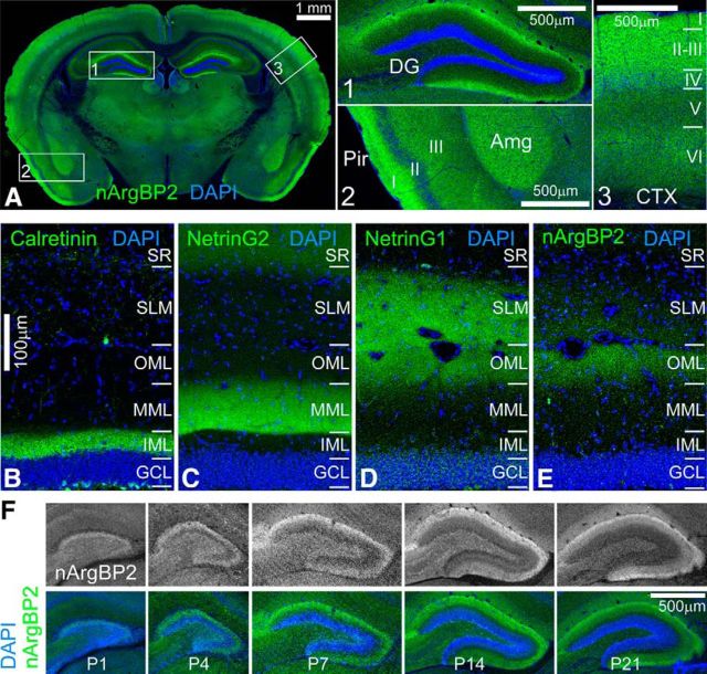
Figure 2.
Expression patterns of nArgBP2 in the mouse brain. A, A coronal section shows expression patterns of nArgBP2 in the mouse brain. Boxes 1–3 outline brain regions that are magnified. nArgBP2 is highly expressed in the DG molecular layer (Box 1), the amygdala (Amg) and layer I of piriform cortex (Pir) (Box 2), and also layer I-III of neocortex (Box 3). B–E, Comparison with DG inner molecular layer (IML) marker Calretinin (B), medial molecular layer (MML) marker NetrinG2 (C), and outer molecular layer (OML) marker NetrinG1 (D) reveal that nArgBP2 is enriched in the OML (E). F, nArgBP2 distribution in DG during development from P1 to P21. SR, Stratum radiatum; SLM, stratum lacunosum-moleculare; GCL, granule cell layer.

