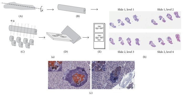
Figure 1.
Schematic illustration of sampling principles for stereological assessment. (a) Formalin fixed pancreas samples were rolled tightly into strips of gauze, infiltrated in paraffin, and cut into 3-4 systematic uniform random tissue slabs with a razor blade fractionator and embedded in one paraffin block with the cut surface down. The blocks were trimmed and four sections for each animal were sampled 300 μm apart and arranged on two glass slides (b), representing a systematic uniform random sample of the whole pancreas. (c) Representative images of pancreatic sections from untreated mice demonstrating β-cells (brown: insulin), non-β-cells (black: glucagon, somatostatin, and pancreatic polypeptide), unstained endocrine cells (arrow), and surrounding immune cells (double arrow: hematoxylin). Scale bars = 400 μm.

