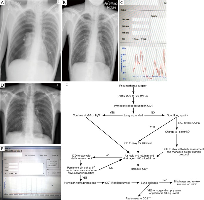
Figure 3.
An example of a pneumothorax patient managed initially with a traditional chest drain. (A) Right pneumothorax; (B) chest drain in situ connected to a traditional one-bottle system; (C) after 5 days, connection to DDS showed persistent airleak for 24 hours with the unlikely hood of resolving; (D) post-VATS pleurodesis CXR with no evidence of pneumothorax; (E) 48 hours of flat line on DDS before drain removal; (F) flow chart with recommendations of chest tube management following pneumothorax surgery connected with DDS. *, although the management in general does not differ between primary and secondary pneumothorax it is imperative to adjust the suction depending on the quality of the underlying lung parenchyma; **, in cases of significant emphysema, drains should be removed when there is no excessive swing on the tubing; ***, the vast majority of patients will experience significant lung collapse when there is a large air leak on the background of severe emphysema. Suction should be low and never exceed −8 cmH2O although lower settings might be necessary in the presence of large leak. An individual approach is necessary to achieve best balance between adequate suction without promoting an air leak. DDS, Digital Drain System. VATS, video assisted thoracoscopic surgery; ICD, intercostal drain; CXR, chest X-rays.

