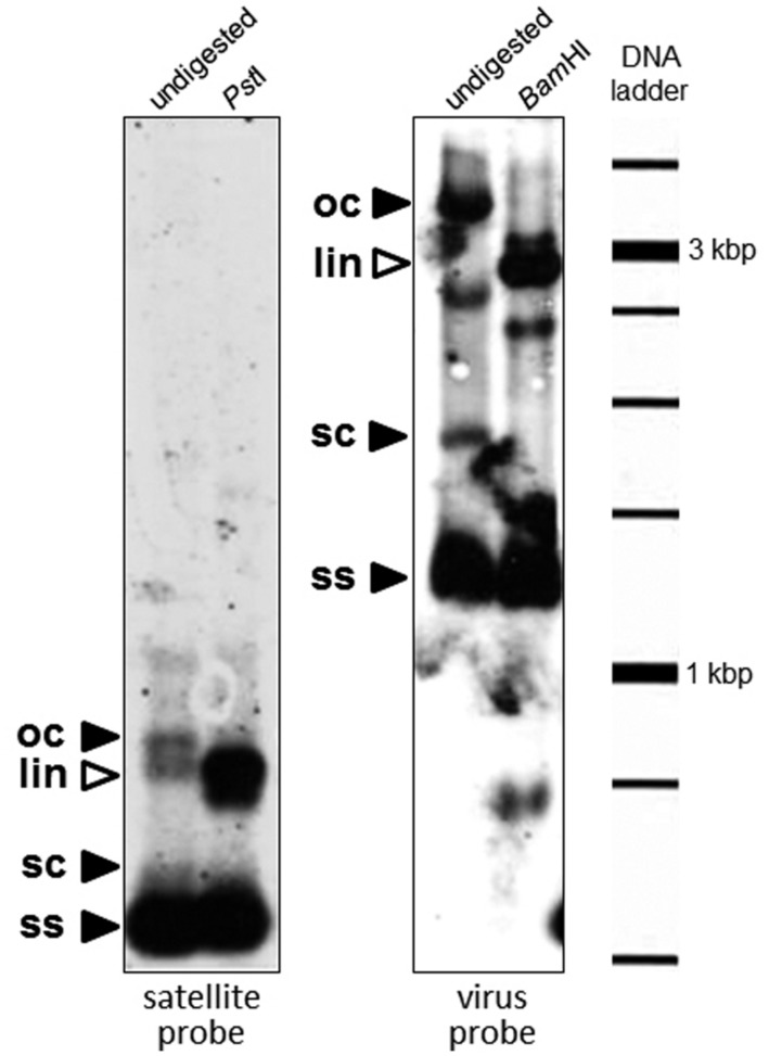
FIGURE 4.
Southern blot analysis to detect the presence of DNA satellites in a sweepovirus-infected sweet potato sample (B3). Total DNA (∼1 μg) was separated on 0.8% agarose gel electrophoresis in TAE, transferred to a positively charged nylon membrane and hybridized with digoxigenin-labeled probes synthesized by PCR from clone SBG53 (satellite, left panel) and the sweet potato leaf curl virus (SPLCV) isolate present in that sample (virus, right panel). Total DNA was digested (right part of each panel) with a restriction enzyme which recognizes a unique site in the satellite (PstI) and SPLCV (BamHI). The positions of open circular (oc), supercoiled (sc), and single-stranded (ss) DNA forms are indicated with black arrowheads. The positions of linear (lin) DNA forms are indicated with white arrowheads. Mobility of the size marker (1 kb DNA ladder) is given in the right margin.

