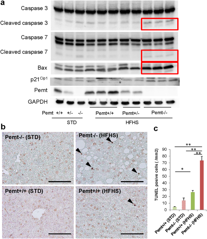
Figure 4. Apoptosis of hepatocytes in Pemt+/+, Pemt+/− and Pemt−/− mice at 25 weeks of age.
(a) Western blot analyses of liver tissue samples for pro-apoptotic molecules, caspases 3 and 7, Bax, and p21Cip1. Cleaved caspase 3 (17 and 19 kDa), cleaved caspase 7 (20 kDa) and Bax prominently up-regulate in Pemt−/− mice fed high fat-high sucrose diet (red squares). (b) TUNEL staining of the liver. TUNEL positive cells are shown by arrow heads. c. TUNEL-positive apoptotic cells/mm2. The apoptotic cells increase in Pemt−/− mice compared to Pemt+/+ mice under high fat-high sucrose diet. All data are presented as mean ± S.E. n = 3–6. *p < 0.05, **p < 0.01.

