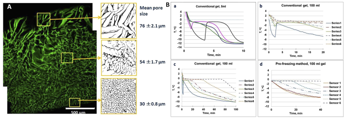
Figure 2.
(A) Confocal laser scanning microscopy (CLSM) image of a cross section of gelatin hydrogel prepared in a petri dish. The freezing started from the bottom of the petri dish where small pores formed, while larger pores formed at the contact with air (top). The scale bar is 500 mm. (B) Temperature profiles measured in HEMA cryogel samples: a) four different samples 5 ml each, b) and c) 100 ml sample prepared using conventional method; and d) 100 ml sample after pre-freezing.

