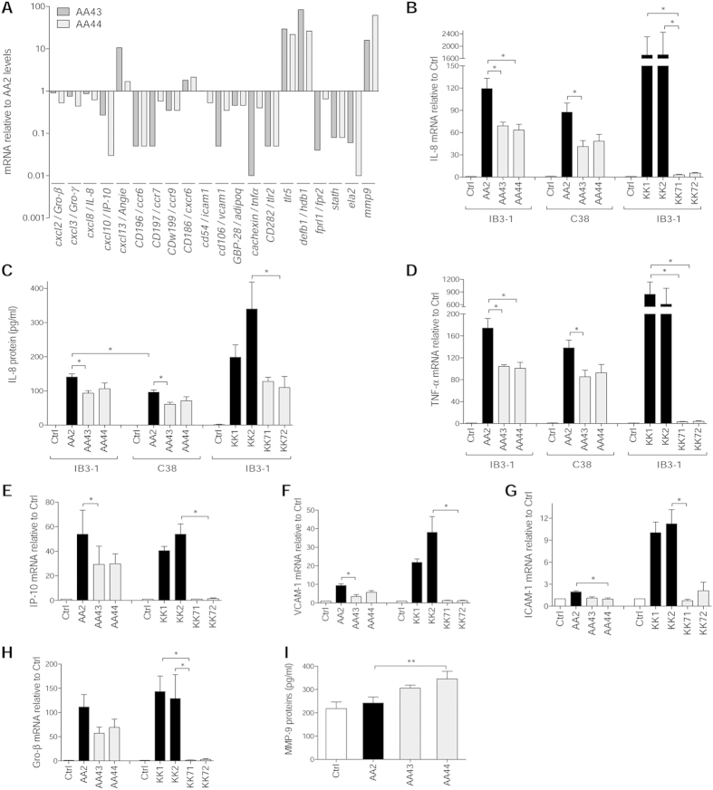
Figure 4. Expression/release of markers of inflammation and tissue damage in cell lines after infection with P. aeruginosa phenotypic variants.
Bronchial epithelial CF cells IB3-1 were infected for 4 hours with P. aeruginosa AA2-AA43-AA44 isolates, RNA extracted and retrotranscribed, and macroarray conducted. (A) Genes expression is expressed after normalization on expression induced by AA2. Validation of gene expression was performed by real time PCR in IB3-1 (B,D–H) and isogenic non-CF cells C38 (B,D) after infection with AA2, AA43, AA44, KK1, KK2, KK71 and KK72. (C) Validation of IL-8 protein release was performed by ELISA in culture medium of IB3-1 and C38 after infection with isolates mentioned above. (I) Macrophagic-like cells THP-1 were infected for with P. aeruginosa AA2, AA43 and AA44 isolates (MOI 1), and MMP-9 release was measured in the culture supernatants by ELISA. Values represent the mean ± SEM. The data are pooled from at least three independent experiments. Statistical significance is indicated: *p < 0.05, **p < 0.01.

