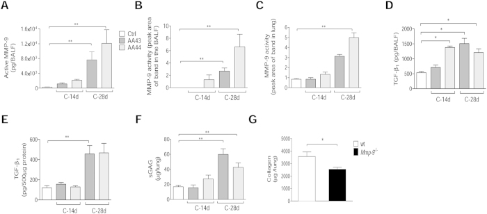
Figure 6. Tissue damage markers, including MMP-9, in murine models after P. aeruginosa infection.
C57Bl/6NCrlBR mice were infected with 1 to 2 × 10⁶ CFU/lung of CF-adapted isolates embedded in agar beads for 14 and 28 days. Levels of MMP-9 protein (A) in BALF by ELISA, MMP-9 activity (B) in BALF and (C) lung homogenate by zymography, TGF-β1 (D) in BALF and (E) lung homogenate by Bioplex and sGAG (F) in lung homogenate by a dye-binding colorimetric assay were measured after 14 and 28 days of chronic lung infection. Values represent the mean ± SEM. The data are pooled from at least two independent experiments (n = 3–12). G) B6.FVB(Cg)-Mmp9tm1Tvu/J and congenic mice were infected with 2 × 10⁶ CFU/lung of AA43 strain embedded in agar beads. Collagen levels were evaluated by a dye-binding assay in lung homogenate after 28 days of chronic lung infection with the P. aeruginosa CF-adapted isolate AA43. Values are represented as mean ± SEM. The data derive from one experiment (n = 6–7). Statistical significance is indicated: *p < 0.05, **p < 0.01.

