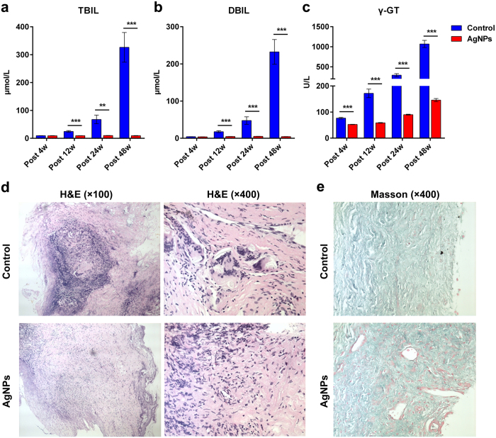
Figure 6. AgNP biliary stent maintained good biliary status in vivo.
To monitor biliary status in real time, blood samples were collected serially via an ear vein at 4, 12, 24 and 48 weeks after biliary stent implantation to test liver function in the other twenty-four pigs for each group. Plasma levels of (a) total bilirubin (TBIL) and (b) direct bilirubin (DBIL) and (c) γ-L-glutamyl dipeptide (γ-GT) were compared between the AgNP group and the control group at different time points post-operation. **p < 0.01; ***p < 0.001. (d) The histology of bile duct anastomosis in the AgNP group and control group was observed by haematoxylin and eosin (H&E) staining. Magnification: 100×; 400×. (e) The collagen fibre (staining green) and myofibre (staining red) of bile duct anastomosis in the AgNPs group and control group were observed using Masson’s trichrome staining. Magnification: 400×.

