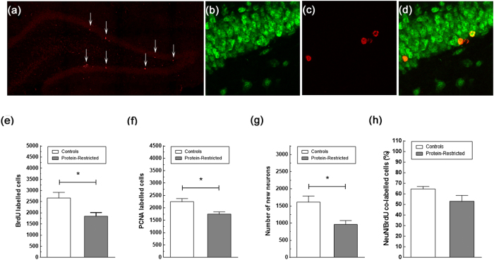
Figure 5. Impact of early protein-restriction on hippocampal neurogenesis at adulthood.
Representative confocal images of the dentate gyrus stained for PCNA (a), NeuN (b), BrdU (c), and double stained for BrdU and the neuronal marker NeuN (d) in controls rats. Arrows in (a) point to PCNA immuno-positive cells. Cells double-labeled with BrdU and NeuN were counted as new neurons. Data correspond to the mean ± SEM of the total number of BrdU (e), PCNA (f) and double labeled cells (g), and to the percentage of BrdU/NeuN stained cells in relation to the total number of BrdU positive cells (h). Note that, though naïve PR rats exhibit a reduced number of BrdU-labeled cells co-expressing the neuronal marker NeuN and, therefore, of new neurons, there are no differences between control and PR rats in the proportion of BrdU/NeuN-labeled cells *p < 0.05, Student’s t-test with n = 5–8 animals per group.

