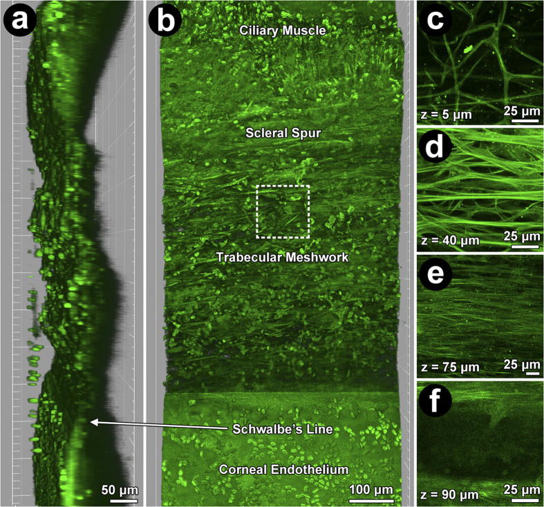
Figure 1. 2-photon excitation fluorescence imaging (TPEF) of human trabecular meshwork (TM) showing autofluorescent extracellular matrix (ECM) and Hoechst-33342-labeled nuclei (green ovals) at low (220X) and high magnification (756X).
(a) Orthogonal view: TM between ciliary muscle and scleral spur (posterior; top), and Schwalbe’s line and Descemet’s membrane (anterior; bottom). (b) En face view: TM cells and autofluorescent fibers between ciliary muscle and cornea. Dashed box: region of high magnification images (756X) of uveal (c; UM), corneoscleral (d; CSM), juxtacanalicular meshwork (e; JCT) and Schlemm’s canal (f; SC). (c): UM (mean ± SD thickness: 25 ± 14 μm) with slender branching beams separated by wide gaps (diameters >40 μm). (d): CSM autofluorescent plate-like beams (than UM) with smaller pores (diameters <40 μm) was 25–65 μm (~40 μm average thickness) from UM inner surface. (e): JCT arrays of fine autofluorescent fibers orientated parallel to SC, 65–75 μm (~10 μm average thickness) from UM inner surface. (f): Autofluorescence signal void of SC ~75 μm from inner UM surface.

