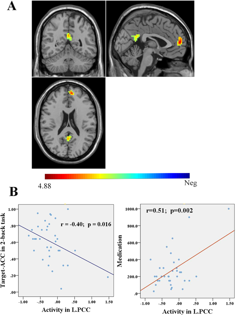
Figure 3. Differences of DMN activity in the 2-back working memory task between healthy controls and schizophrenia patients with impaired cognitive function.
(A) shows greater activity (decreased suppression) in the left mPFC and PCC in schizophrenia patients with impaired cognitive function compared to health controls (cluster p < 0.05 with FWE correction); (B) shows the significant correlations of the left PCC activity with Target-accuracy of 2-back task and with medication dosage in patients. mPFC, medial prefrontal cortex; PCC, posterior cingulated cortex. The color bar represents the T values.

