
Figure 2.
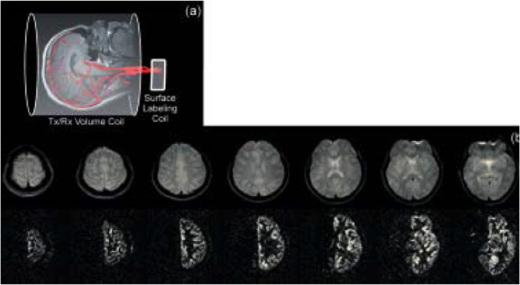
(a) An alternative option that excludes the need of the oblique labeling plane is to place the labeling RF coil right over one of the carotids. (b) Multi-slice axial control (top) and subtraction (bottom) images created using a unilateral carotid labeling RF coil and spin echo EPI (TR/TE/labeling period/post-labeling delay = 4s/22ms/3s/0.5s). As expected in the normal brain, perfusion from one carotid artery supplies only the ipsilateral hemisphere. The excellent subtraction of the contralateral hemisphere serves as a demonstration of the elimination of magnetization transfer effects. This method requires repositioning the coil to selectively label the contra lateral carotid. (Reproduced by permission of Zaharchuk et al (15)).