
Figure 4.
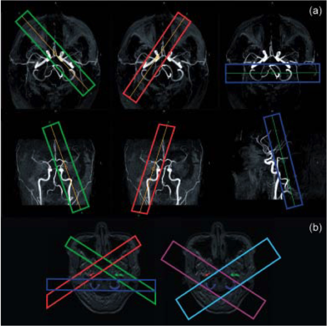
(a) Planning of the respective labeling of the left ICA (green), right ICA (red), and posterior circulation (blue) on the MIPs of the circle of Willis of a healthy volunteer. (Reproduced by permission of Golay et al (25)) (b) Planning of the labeling scheme used in the dual vessel approach, in which two acquisitions (light blue and purple), both including the posterior circulation, replace the three acquisitions (red, green and blue) from the previous method. (Courtesy of Dr. Xavier Golay).