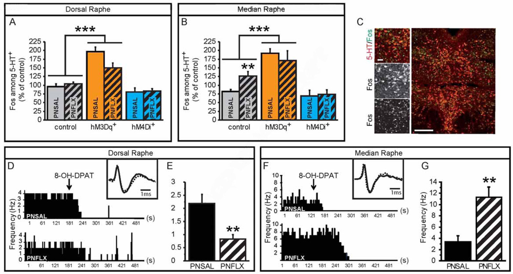
Figure 6. Altered balance between DR and MR 5-HTergic neuronal activity in PNFLX mice.
(A–C) Fos and 5-HT immunostaining on hM3Dq+ and hM4Di+ mice and controls in DR (A) and MR (B) demonstrates an increase of in %Fos+/5-HT+ neurons in hM3Dq+ mice when compared to control animals. Moreover, PNFLX control animals display an increase in %Fos+/5-HT+ neurons in the MR (B), but not the DR (A) when compared to PNSAL control animals. n = 3–9 per genotype, post-natal treatment and region.
(D–G) The anatomical location of in vivo electrophysiological recordings of putative 5-HTergic raphe neurons was validated at the end of each recording session through responsiveness to 5-HT1A receptor mediated inhibition of neuronal firing, achieved by 8-OH-DPAT administration (ip, 1 mg/kg) (D, F). Insets show examples of single extracellularly recorded spikes from PNSAL (plain line) and PNFLX (dashed lines) animals. The firing frequency of putative 5-HTergic neurons of PNFLX mice were reduced in the DR (E) but increased in the MR (G) when compared to PNSAL animals. n = 8–9 animals.
* p < 0.05; ** p < 0.01; *** p < 0.005. Scale bars are 20µm and 100µm. DR: dorsal raphé, MR: median raphé.

