
Fig. 4.
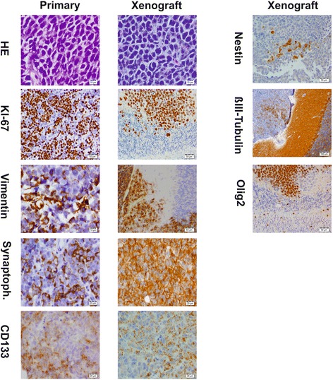
Immunohistological evaluation of the original tumor and xenografted infratentorial tumor. Selected markers are shown in comparison. For some markers, there was not sufficient material left from the primary tumor. All specimens were formaldehyde fixed and paraffin embedded prior to histopathological evaluation