
Fig. 2.
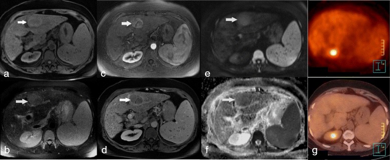
Moderately differentiated (grade 2) HCC in a patient with hepatitis B-induced cirrhosis, showing restricted ADC on DWI-MRI and no significant uptake on FDG-PET/CT. MR imaging showed a left lobe HCC with high T1 (a, arrow), high T2 (b, arrow) signal intensity, with arterial enhancement (c, arrow) followed by washout (d, arrow). The HCC showed high signal intensity on DWI image (b = 800 s/mm2; e, arrow) consistent with low ADC (1.26 × 10⁻³ mm2/s) on the ADC map (f, arrow). The lesion showed no significant FDG uptake on FDG-PET/CT (g)