Fig. 7.

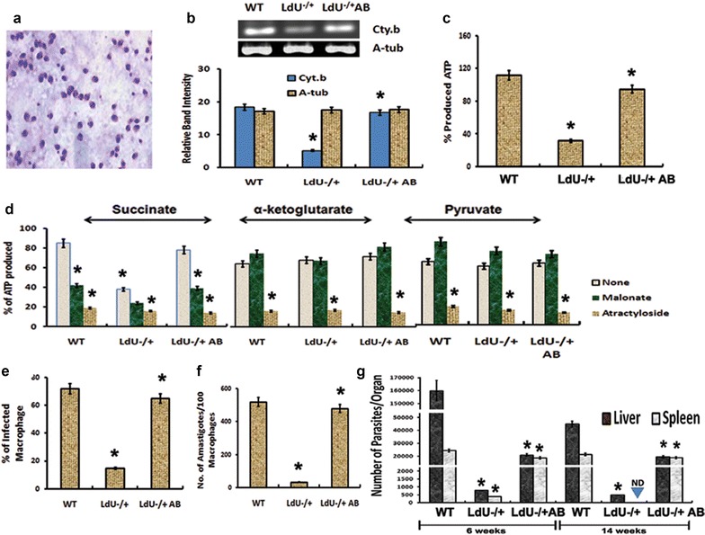
Effect of UMSBP deletion on amastigote survival both in macrophages and in mice by interfering with electron transport chain, oxidative phosphorylation and ATP generation. a Image of isolated amastigotes from infected macrophages. b mRNA level of Cyt. b in WT, LdU−/+ and LdU−/+AB amastigotes by semiquantitative-RT-PCR. c % of ATP production in mitochondria of WT, LdU−/+ and LdU−/+AB amastigotes. d ATP production by mitochondria from the indicated intracellular amastigotes was evaluated for three substrates (succinate, α-ketoglutarate and pyruvate, each indicating a separate mitochondrial pathway). The tested substrate is indicated at top. Addition of these compounds to the samples or no inhibitor (none) is indicated by the bar graphs shading according to the legend. ATP production in mitochondria of WT cells without addition of malonate or attractyloside was set to 100 % for each substrate. e–f Viability of amastigotes inside human macrophages after knocking out LdUMSBP as well as after complementation of WT UMSBP in LdU−/+ parasites (LdU−/+AB). e Graph showing the numbers of infected macrophages for the indicated parasites. f Graph showing the numbers of amastigotes in infected macrophages for the indicated parasites. g Survival of WT, LdU−/+ and LdU−/+AB parasites in BALB/c mice. Parasite burdens were analyzed at 6 or 14 weeks post-infection by serial dilution as described in “Methods” section. Asterisk denotes that the data are significant (p ≤ 0.001)
