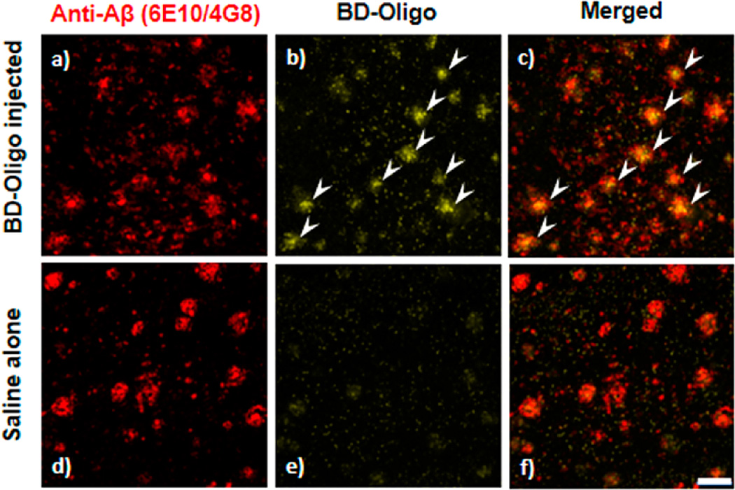
Figure 4.
Ex vivo binding of BD-Oligo in 18 month old AD mouse brains. (a, b, and c) Fluorescence in the APP/PS1 mouse brain injected with BD-Oligo using the channel for 6E10/4G8 labeling, BD-Oligo labeling, and the merged image, respectively. BD-Oligo fluorescence was present in the brain 24 h after an ip injection of BD-Oligo (see b), which colocalized with the Aβ labeling (see c). Arrows indicate plaques with colocalization. (d, e, and f) Fluorescence in the APP/PS1 mouse brain injected saline alone using the channel for 6E10/4G8 labeling, BD-Oligo labeling, and the merged image, respectively. There are no plaques seen in the BD-Oligo channel in the control saline-injected mice, indicating the specificity of the BD-Oligo oligomer labeling. Scale bar 100 µm.

