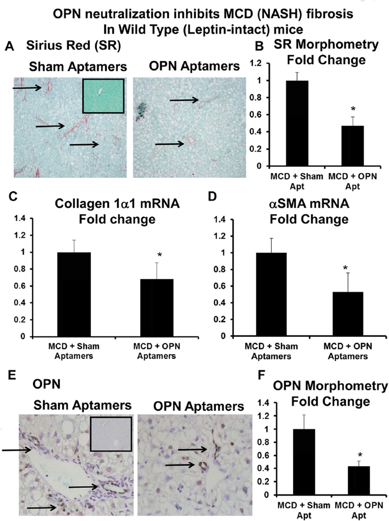
Fig. 6.
OPN neutralization inhibits diet-induced NASH fibrosis in leptin-intact mice. Leptin-intact (WT) mice were fed control chow or the MCD diet for 6 weeks, in the presence of sham or OPN-aptamers. Mice were sacrificed 24 h after final dose of aptamers, and livers analyzed. (A) Representative Sirius-red staining; black arrows indicate Sirius-red stained fibrils. Insert shows Sirius-red staining from a control-fed mouse. (B) Sirius-red morphometry. (C) Collagen 1α1 mRNA. (D) αSMA mRNA. Results are expressed as fold changes relative to sham-aptamer treated mice, and graphed as mean ± SEM. *p < 0.05 vs. sham-aptamer treated mice. (E) Representative OPN staining; black arrows indicated OPN-positive cells. Insert shows OPN staining from control-fed mouse. (F) OPN morphometry.

