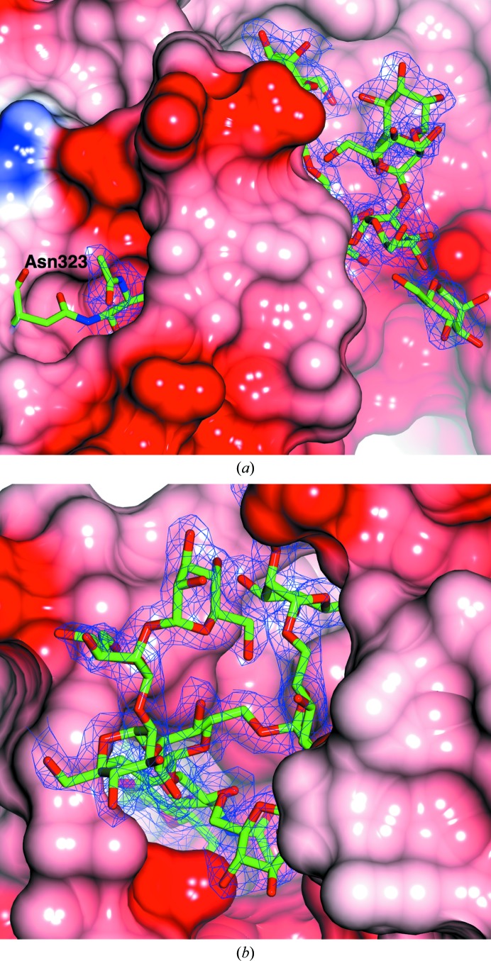
Figure 3.
The electron density for the glycosylation tree attached to Asn323 in AfβG shown from two different perspectives. In (a) the first part of the tree is buried within a pocket of the protein. In (b) Asn323 is at the base of the pocket. There is well ordered density for all of the sugars. The maximum-likelihood map was contoured at the 1σ level.

