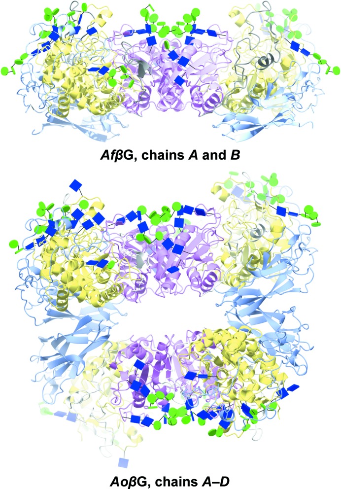
Figure 4.
Three-dimensional fold, domain organization and asymmetric unit packing of AfβG and AoβG. Both enzymes have three domains (A, yellow; B, pink; C, light blue), with a dimer being the preferred biological arrangement. AoβG has two dimers in the asymmetric unit, with all of the sugars facing opposite sides. The sugars are shown as glycoblocks, with blue squares for N-acetyl-β-d-glucosamine and green circles for d-mannopyranose.

