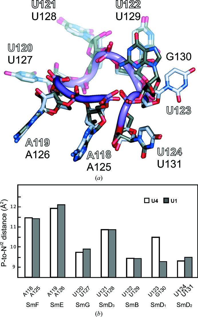
Figure 9.
Comparison of the U4 and U1 Sm-site heptads. (a) The U4 and U1 Sm-site heptads are superimposed and viewed from the flat face at a glancing angle. The only notable difference occurs at the sixth nucleotide, where the U123 base of U4 snRNA is bound in the pocket of SmD1, while the G130 base of U1 snRNA lies outside the central hole. The G130 base partially covers the base of U129, which is stacked on the face near G130 with His37 in L3 of SmB. Thus, shielding of UV irradiation by G130 is likely to have prevented the cross-linking of U1 snRNA to L3 of SmB (Urlaub et al., 2001 ▸). (b) Distances from the P atom of the Sm-site nucleotide to the Nδ2 atom of the invariant Asn in its binding pocket. The P-to-Nδ2 distance measures the RNA backbone position relative to the depth of the pocket, where the base is hydrogen-bonded to Nδ2. The distances are NCS-averaged with standard deviations of 0.04–0.10 Å at each position. The distances are closely similar between the U4 and U1 Sm sites in all except the sixth pocket and they are longer for A than for U. In the sixth pocket, G130 of U1 snRNA, which shows a shorter P-to-Nδ2 distance than U123 of U4 snRNA, is excluded from the SmD1 pocket.

