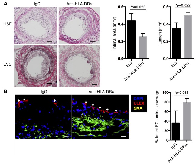
Figure 1. HLA-DR blockade reduces acute T cell–mediated injury to implanted allogeneic vessel segments in vivo.
(A and B) In an MHC-mismatched model of arterial rejection by allogeneic T cells (n = 6 per group), blockade of class II MHC by anti–HLA-DRα F(ab)′2 fragment reduces intimal area expansion and increases lumen area as measured by H&E and EVG staining (scale bar: 50 μm) (A) and reduces disruption of EC lining the vessel lumen, a hallmark of intimal arteritis/endothelialitis, as measured by percent of circumferential coverage (scale bar: 20 μm) (B), both at 21 days. Arrows indicate areas of intact endothelial lining. *P < 0.05, 2-tailed Student’s t test. EVG, Elastia-van Gieson; MHC, major histocompatibility complex; EC, endothelial cells.

