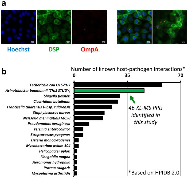
Figure 6. Ab5075 host-pathogen interactions.
(a) Confocal micrographs of Ab5075-infected H292 cells stained with primary antibodies against desmoplakin (green) and OmpA (red). DNA was stained with Hoechst 33342 (blue). Confocal images and overlaid images from replicate analyses at a minimal thickness of 1.1μm, scale bars represent 10μm. (b) The 46 host-pathogen interactions identified in this study (green bar) compared to the number of interspecies interactions identified for other bacterial species (HPIDB 2.0 database) [60]. If substrains were present in the database, the strain with the highest number of interactions was shown. Due to scale, HPIDB 2.0 host pathogen interactions for Yersinia pestis (n=4018), Bacillus anthracis (n=3061), and Francisella tularensis subsp. tularensis SCHU S4 (n=1346) were not shown.

