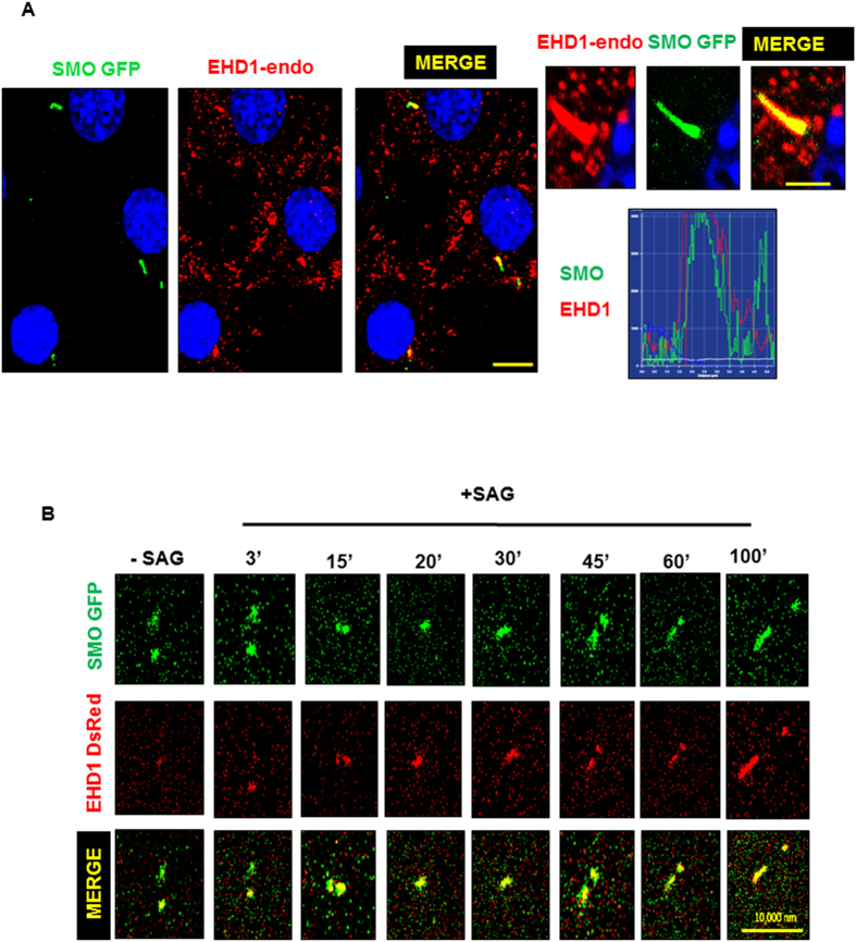
Figure 11. EHD1 co-localizes and co-traffics with SMO to the cilia upon SHH pathway stimulation.
(A) WT NIH3T3 cells stably expressing SMO-GFP were starved in low serum media for 24 hours and stimulated with SAG in starvation media for another 24 hours. Under these conditions EHD1 was seen to co-localize with SMO in the primary cilia. A profile scan for SMO and EHD1 in the primary cilia merged panel is shown. Under these conditions, EHD1 was seen to traffic to the cilia of 60% cells studied. (B) WT NIH3T3 cells stably expressing SMO-GFP and transiently expressing EHD1-DsRed were starved in low serum media for 24 hours and stimulated with SAG immediately before starting live imaging of Smoothened and EHD1.As reported in earlier studies, Smoothened was found in preciliary vesicles under non-stimulated conditions but upon SHH pathway activations, EHD1 was seen to associate with Smoothened vesicles and co-traffic with SMO into the primary cilia.

