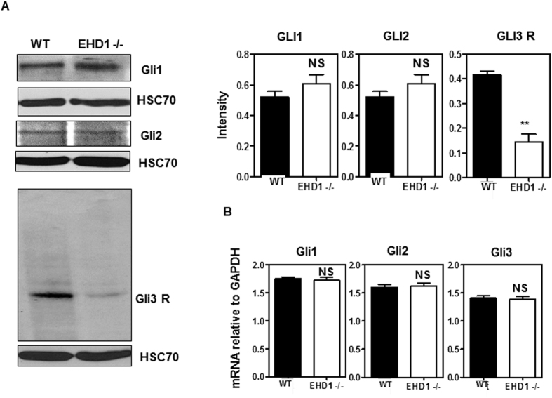
Figure 6. Ehd1-null embryos reveal features indicative of increased SHH signaling.
(A) 40 μg aliquots of whole embryo lysate protein from pooled E9.5 WT and Ehd1-null embryos were separated using 8% SDS-PAGE and immunoblotted using antibodies against GLI1, GLI2 and GLI3. HSC-70 is the loading control. The blot is a representative one from three individual experiments. Data from multiple experiments are presented as mean ± S.E.M. (error bars, n = 3) with levels of expression normalized to HSC70 expression in each experiment. The membrane for GLI1 was serially stripped and reprobed with GLI2, followed by Hsc70 antibodies. A separate membrane was probed for GLI3.GLI3 repressor levels are markedly reduced in Ehd1-null embryos (P < 0.05) whereas GLI1 and GLI2 expression are unchanged between Ehd1-null and WT control littermates. Unpaired t test; n = 3 for each condition. Full-length blots/gels are presented in Supplementary Figure S9. (B) Relative mRNA levels of Gli1, Gli2 and Gli3 in the Ehd1-null and WT embryos measured by qRT-PCR analysis. Gli1, Gli2 and Gli3 mRNA levels remain comparable between Ehd1-null and WT embryos. Unpaired t test; n = 3 for each condition.

