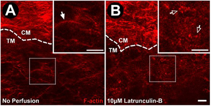
Figure 8. Effect of Lat-B on aqueous drainage tissue F-actin following live mouse drug delivery.
(A) F-actin labeling (red) of unperfused control eye (n = 3 mice). Ciliary muscle (CM) F-actin labeling is brighter and denser compared with adjacent trabecular meshwork (TM). Cortical F-actin organized as a curvilinear network is prevalent without punctate F-actin. (B) F-actin after Lat-B (10 μM, n = 3) delivery in live mice. The curvilinear F-actin network was absent but punctate F-actin was prominent. Closed arrows: Cortical F-actin organized as curvilinear network. Open arrows: Punctate F-actin. Dashed lines: CM-TM border. Insets: 2× magnification of sample region in the TM. Semi-transparent boxes: indicate region of TM selected for magnification in inset. Scale bar, 25 μm.

