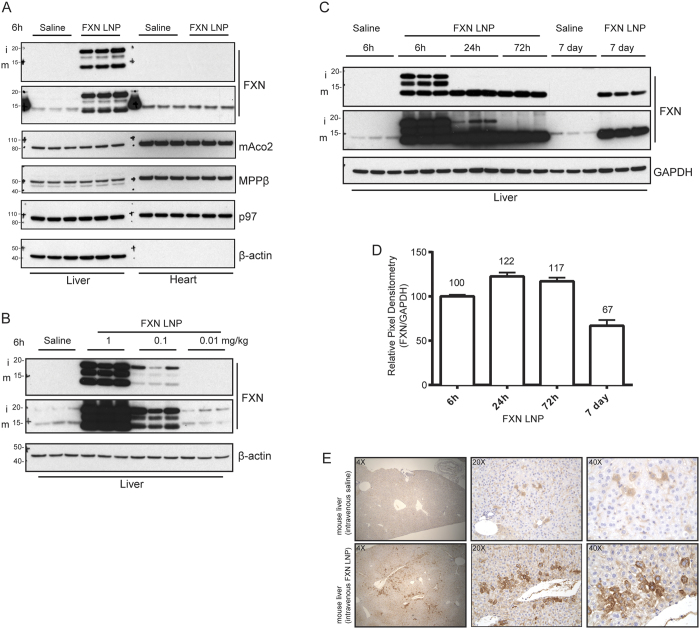
Figure 2. FXN mRNA delivered using LNP vehicle in vivo is efficiently translated and processed in liver.
(A) Mice were injected IV with FXN LNPs or saline solution as control (3 animals per group). Tissues, including liver and heart, were collected 6 h post-injection and frozen before preparation of homogenates for western analysis. Immunoblotting was carried out with the indicated antibodies. Two antibodies targeting FXN were used to confirm the immunopositive signal corresponding to FXN (upper panel: abcam ab110328; lower panel: Proteintech 14147-01-AP). p97 and β-actin were used as lane-loading controls. (B) Various concentrations of FXN LNP were tested by IV administration. Lysates derived from livers 6 h after IV injection of LNP or saline solution (3 animals per group) were prepared and analyzed by immunoblotting with the indicated antibodies. Two antibodies targeting FXN were used to confirm the immunopositive signal corresponding to FXN (upper panel: abcam ab110328; lower panel: Proteintech 14147-01-AP). (C) Mice were injected IV with FXN LNP or saline solution and animals were necropsied at different timepoints post-LNP administration. Liver homogenates were prepared and analyzed by western blotting with the indicated antibodies. Two antibodies targeting FXN were used to confirm the immunopositive signal corresponding to FXN (upper panel: abcam ab110328; lower panel: Proteintech 14147-01-AP). (D) Pixel densitometry analysis of the FXN LNP –derived mFXN immunopositive signals at 6 h, 24 h, 72 h, and 7 days post-administration. Densitometries were normalized relative to levels at 6 h. (E) Anti-FXN IHC staining of mouse livers 24 h after administration of FXN LNPs or saline solution. Three magnifications (4×, 20×, and 40×) of the same region are shown.

