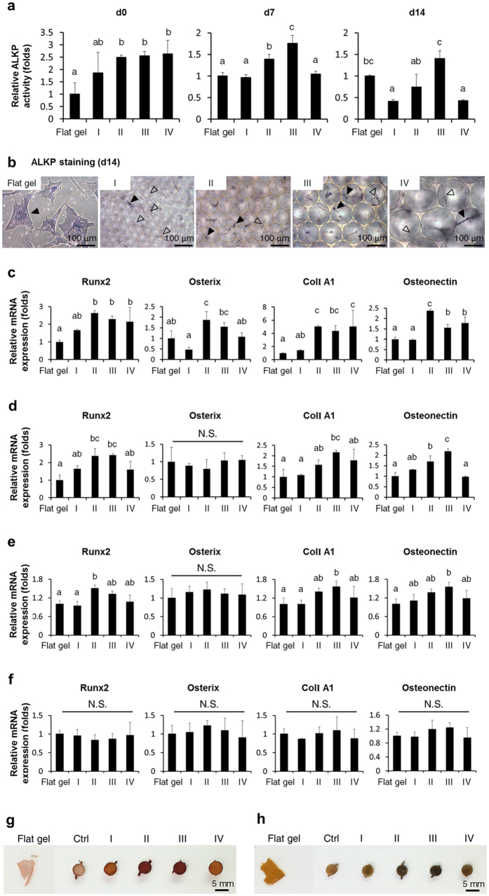
Figure 4. Osteogenic differentiation of MSCs in the 3D scaffolds (Groups I, II, III, and IV) and on the 2D flat gel.
(a) Analysis and quantification of ALKP activity of differentiating MSCs in the 3D scaffolds or on the 2D flat gel at 0, 7, and 14 days of culturing in osteogenic medium. (b) ALKP staining of differentiating MSCs in the 3D scaffolds or on the 2D flat gel at 14 days of culturing in osteogenic medium. ALKP positive cells were stained bluish-purple. Solid arrows indicated positively stained cells and hollow arrows indicated negative staining. Osteoblast-related gene expressions of differentiating MSCs in the 3D scaffolds or on the 2D flat gel were determined by qPCR after (c) 1 day of culturing in the maintenance medium and (d) 7, (e) 14, and (f) 21 days of culturing in the osteogenic medium. Data were represented as mean ± SD of the ratios of the 3D groups to the flat gel group, n = 3. Groups with different letters were significantly different, whereas groups with same letters were not; p < 0.05. N.S., no significance. (g) Alizarin red S and (h) von Kossa staining of differentiating MSCs in the 3D scaffolds or on the 2D flat gel at 28 days of culturing in the osteogenic medium. Ctrl represented control group with 3D scaffold only. Alizarin red S colored calcium deposits were stained red. Von Kossa staining demonstrated blackened in calcium salts.

